Improved Image Quality and Resolution for all ZEISS Laser Scanning Microscopes
ZEISS LSM Plus and ZEISS Airyscan Joint Deconvolution introduced
Today, ZEISS introduces two new software functionalities: With ZEISS LSM Plus and ZEISS
Maximizing recovery requires improvements in efficiency
Laser scanning microscopes (LSMs) are the multifunctional high-end microscopes in every imaging facility, valued as much for their instant, high-quality imaging of optical sections as for their ease of use and application flexibility. ZEISS LSMs provide a wide range of detection modes with highest sensitivity and spectral flexibility for multi-fluorescent experiments. ZEISS LSM Plus and ZEISS Airyscan Joint Deconvolution significantly improve the data quality in confocal imaging. This enhancement is independent of the chosen detector, imaging mode, and emission range.
ZEISS LSM Plus: Improving the whole confocal experience
The linear Wiener filter deconvolution of the new ZEISS LSM Plus option is based on the time-tested super-resolution processing of ZEISS Airyscan. It can be applied to data of any confocal imaging mode - including spectral, near infrared and multiphoton microscopy - with next to no interaction, ensuring reliable quantitative results. Users benefit from an enhanced signal-to-noise ratio at high acquisition speed and low laser power - particularly useful for live cell imaging of delicate samples. For samples that are not too light sensitive and therefore allow the pinhole of the LSM to be closed, more spatial information and even greater resolution enhancement can be obtained.
ZEISS Airyscan Joint Deconvolution: 32 views mean more information
ZEISS Airyscan 2 is an area detector with 32 circularly arranged detection elements. Each of
these acts as a small pinhole with a slightly different view on the sample, providing additional spatial information. With the introduction of ZEISS Airyscan Joint Deconvolution, this additional spatial information can now be leveraged to reduce the distance that can be resolved between two points down to 90 nm. Super-resolution experiments benefit from an improved separation of single or multiple labels. Users immediately recognize the significance for their research: "When we imaged endoplasmic reticulum and mitochondria and saw their very fine details after applying ZEISS Airyscan Joint Deconvolution, we thought this is just super cool. The new option could be integrated within our imaging protocols very easily. We were amazed at how quickly the images were processed, which helped us make decisions while we were still imaging", says Dr. Kelly Subramanian, Post-Doctoral Scholar, Department of Molecular and Cellular Biology at UC Davis,
USA.
Users benefit from ease of use
These two new options enable users to reliably gather more information for all confocal
experiments while increasing productivity. Particularly, imaging facilities with multiple users
benefit from seamlessly integrated, easy-to-apply improvements - without any expert knowledge or extra effort. The existing ZEISS LSM 900, including ZEISS Celldiscoverer 7, and ZEISS LSM 980 microscope systems can be upgraded accordingly.
Featured Product
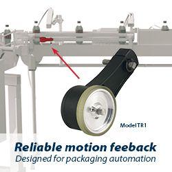
Model TR1 Tru-Trac
The Model TR1 Tru-Trac® linear measurement solution is a versatile option for tracking velocity, position, or distance over a wide variety of surfaces. An integrated encoder, measuring wheel, and spring-loaded torsion arm in one, compact unit, the Model TR1 is easy to install. The spring-loaded torsion arm offers adjustable torsion load, allowing the Model TR1 to be mounted in almost any orientation - even upside-down. The threaded shaft on the pivot axis is field reversible, providing mounting access from either side. With operating speeds up to 3000 feet per minute, a wide variety of configuration options - including multiple wheel material options - and a housing made from a durable, conductive composite material that minimizes static buildup, the Model TR1 Tru-Trac® is the ideal solution for countless applications.